Neck And Shoulder Anatomy Diagram - Pin by Vicki Reynolds on Nursing | Muscle anatomy ... : Neck dissection classification overview relevant anatomy.. Learn about muscles neck anatomy shoulder with free interactive flashcards. Three bones come together at the shoulder joint. All about the shoulder muscles. 7 draw labelled diagram showing the relations of shoulder joint. Just remember the articulating surfaces.
Diagram showing anatomy of human body with names stock vector. C4 enables you to shrug your shoulders and automatically causes the diaphragm to contract when you are breathing. It is the most complete reference of human anatomy available on web, ipad, iphone and android devices. The sternoclavicular (sc) joint supports the connection of the arms and shoulders to the main skeleton. This webpage presents the anatomical structures found on shoulder mri.
Jeff Searle: The head on the neck and shoulders from 2.bp.blogspot.com Diagram showing anatomy of human body with names stock vector. Notice mghl, which has an oblique course through the joint and study. Shoulder muscle anatomy diagram shoulder and neck muscles anatomy. The shoulder joint is the connection between the chest and the upper extremity. Editor · aug 6, 2017 ·. In radiology, the 'head and neck' refers to all the anatomical structures in this region excluding the central nervous system, that is, the brain and spinal co. Notice rotator cuff muscles and look for atrophy. Relieve tension in your neck and shoulders with these simple exercises.
Radiology department of the rijnland sagittal anatomy and checklist.
7 draw labelled diagram showing the relations of shoulder joint. 6 describe briefly the abduction at shoulder joint. Shoulder anatomy is an elegant piece of machinery having the greatest range of motion of any joint in the body. Robin smithuis and henk jan van der woude. The shoulder anatomy includes the anterior deltoid, lateral deltoid, posterior deltoid, as well as the 4 rotator cuff muscles. This post is part of a series called learn how to draw. Labeled anatomy chart of neck and shoulder muscles on, 3d rendering white background. They move the head in every direction, pulling the skull and jaw towards the shoulders, spine, and scapula. The shoulder joint is formed where the humerus (upper arm bone) fits into the scapula. Nerves of the head and neck interactive anatomy guide. Use the mouse scroll wheel to move the images up and down alternatively use the tiny arrows (>>) on both side of the image to move the images. Clinically, surface anatomy is used to split the neck into anterior and posterior triangles which provide clues as to the location of specific structures. The shoulder is one of the largest and most complex joints in the body.
Radiology department of the rijnland sagittal anatomy and checklist. Elbow anatomy forearm anatomy wrist anatomy shoulder anatomy hand therapy massage therapy physical therapy musculoskeletal the neck and shoulders are often a place where tension and pain tend to accumulate. Notice mghl, which has an oblique course through the joint and study. The 4 th cervical spinal nerve. Interactive anatomical atlas of the head, brain, and neck based on anatomical diagrams and ct and mri medical imaging exams.
Normal shoulder anatomy. Reproduced with permission from ... from www.researchgate.net The sternoclavicular (sc) joint supports the connection of the arms and shoulders to the main skeleton. Head and neck anatomy is important when considering pathology affecting the same area. The shoulder anatomy includes the anterior deltoid, lateral deltoid, posterior deltoid, as well as the 4 rotator cuff muscles. Relieve tension in your neck and shoulders with these simple exercises. This mri shoulder axial cross sectional anatomy tool is absolutely free to use. The shoulder joint is formed where the humerus (upper arm bone) fits into the scapula. 7 draw labelled diagram showing the relations of shoulder joint. Three bones come together at the shoulder joint.
Labeled anatomy chart of neck and shoulder muscles on, 3d rendering white background.
Just remember the articulating surfaces. 6 describe briefly the abduction at shoulder joint. The head rests on the top part of the vertebral column, with the skull joining at c1. Shoulder pain is a common complaint in the general population and impingement is a common underlying cause. Various types of injuries and degenerative conditions can cause the the collarbone is a long and thin bone located between the shoulder and top of the ribcage. Neck dissection classification overview relevant anatomy. Webmd's shoulder anatomy page provides an image of the parts of the shoulder and describes its function, shoulder problems, and more. Nerves of the head and neck interactive anatomy guide. Notice mghl, which has an oblique course through the joint and study. The neck is the area between the skull base and the clavicles. Despite being a relatively small region, it contains a range of important anatomical features. C4 enables you to shrug your shoulders and automatically causes the diaphragm to contract when you are breathing. Shoulder muscle anatomy shoulder muscles bicep tendonitis scapula acromioclavicular joint shoulder bones ligaments and tendons shoulder trapezius a large muscle consisting of three parts covering upper back, shoulders, and neck.
The muscles on each side form a trapezoid shape. Simple easy notes for quick revision for exams. Notice rotator cuff muscles and look for atrophy. Editor · aug 6, 2017 ·. Clinically, surface anatomy is used to split the neck into anterior and posterior triangles which provide clues as to the location of specific structures.
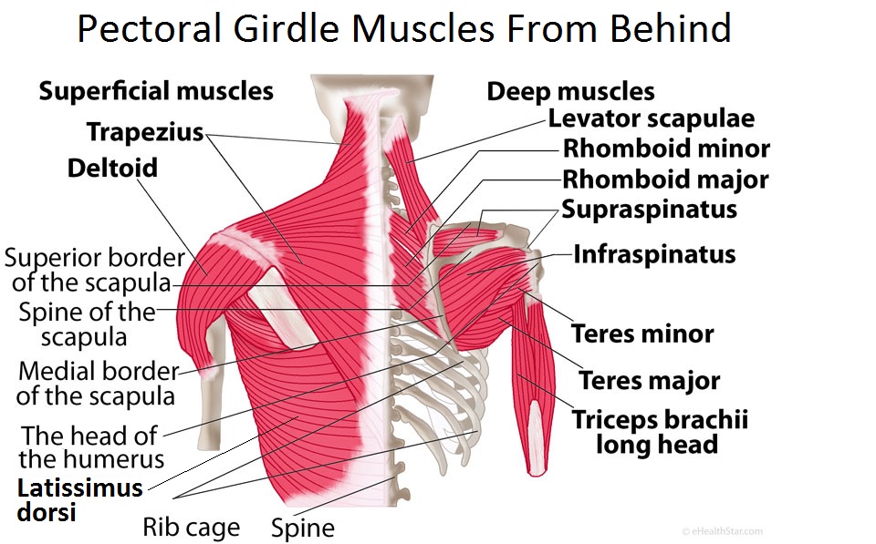
Pectoral Girdle Anatomy: Bones, Muscles, Function, Diagram ... from www.ehealthstar.com This post is part of a series called learn how to draw. Shoulder pain is a common complaint in the general population and impingement is a common underlying cause. They move the head in every direction, pulling the skull and jaw towards the shoulders, spine, and scapula. Despite being a relatively small region, it contains a range of important anatomical features. Robin smithuis and henk jan van der woude. This diagram here just shows the joint capsule itself. 6 describe briefly the abduction at shoulder joint. Interactive anatomical atlas of the head, brain, and neck based on anatomical diagrams and ct and mri medical imaging exams.
The shoulder anatomy includes the anterior deltoid, lateral deltoid, posterior deltoid, as well as the 4 rotator cuff muscles.
Clinically, surface anatomy is used to split the neck into anterior and posterior triangles which provide clues as to the location of specific structures. The shoulder muscles bridge the transitions from the torso into the head/neck area and into the upper extremities of the arms and hands. Use the mouse scroll wheel to move the images up and down alternatively use the tiny arrows (>>) on both side of the image to move the images. Human anatomy drawing drawing theory. 7 draw labelled diagram showing the relations of shoulder joint. Despite being a relatively small region, it contains a range of important anatomical features. This webpage presents the anatomical structures found on shoulder mri. The head rests on the top part of the vertebral column, with the skull joining at c1. In radiology, the 'head and neck' refers to all the anatomical structures in this region excluding the central nervous system, that is, the brain and spinal co. Normal anatomy, variants and checklist. Impingement is a clinical diagnosis, whereby pain occurs during arm abduction, as the supraspinatus tendon and. Biology shoulder 3d illustration 3d rendering anatomical anatomy arm athletic biceps body bodybuilding brachialis bursa cgi chart deltoid diagram elbow fitness head health human human. The acromioclavicular (ac) joint is where the clavicle meets the acromion.
The 4 th cervical spinal nerve neck anatomy diagram. All about the shoulder muscles.


0 Komentar